Welcome to the Amira-Avizo Software Use Case Gallery
Below you will find a collection of use cases of our 3D data visualization and analysis software. These use cases include scientific publications, articles, papers, posters, presentations or even videos that show how Amira-Avizo Software is used to address various scientific and industrial research topics.
Use the Domain selector to filter by main application area, and use the Search box to enter keywords related to specific topics you are interested in.
Three-Dimensional Architecture of Glomerular Endothelial Cells Revealed by FIB-SEM Tomography
Focused-ion beam-scanning electron microscopic (FIB-SEM) tomography enables easier acquisition of a series of ultrastructural, sectional images directly from resin-embedded biological samples. In this study, to clarify the three-dimensional (3D) architecture of glomerular endothelial cells (GEnCs) in adult rats, we manually extracted GEnCs from serial FIB-SEM images and reconstructed them on an Amira reconstruction software. The luminal and basal surface structures were clearly visualized in ... Read more
Yuto Kawasaki, Yasue Hosoyamada, Takayuki Miyaki, Junji Yamaguchi, Soichiro Kakuta, Tatsuo Sakai, and Koichiro Ichimura
The Ebola virus VP40 matrix layer undergoes endosomal disassembly essential for membrane fusion
Ebola viruses (EBOVs) assemble into filamentous virions, whose shape and stability are determined by the matrix viral protein 40 (VP40). Virus entry into host cells occurs via membrane fusion in late endosomes; however, the mechanism of how the remarkably long virions undergo uncoating, including virion disassembly and nucleocapsid release into the cytosol, remains unknown. Here, we investigate the structural architecture of EBOVs entering host cells and discover that the VP40 matrix disassem... Read more
Sophie L Winter, Gonen Golani, Fabio Lolicato, Melina Vallbracht, Keerthihan Thiyagarajah, Samy Sid Ahmed, Christian Lüchtenborg, Oliver T Fackler, Britta Brügger, Thomas Hoenen, Walter Nickel Ulrich S Schwarz, Petr Chlanda
In recent years new methodologies and workflow pipelines for acquiring correlated fluorescence microscopy and volume electron microscopy datasets have been extensively described and made accessible to users of different levels. Post-acquisition image processing, and particularly correlation of the optical and electron data in a single integrated three-dimensional framework can be key for extracting valuable information, especially when imaging large sample volumes such as whole cells or tissu... Read more
Allon Weiner
Cortical bone is permeated by a system of pores, occupied by the blood supply and osteocytes. With ageing, bone mass reduction and disruption of the microstructure are associated with reduced vascular supply. Insight into the regulation of the blood supply to the bone could enhance the understanding of bone strength determinants and fracture healing. Using synchrotron radiation-based computed tomography, the distribution of vascular canals and osteocyte lacunae was assessed in murine cortica... Read more
J.A. Núñez; A. Goring; B. Javaheri; H. Razi; D. Gomez-Nicola; E. Hesse; A.A. Pitsillides; P.J. Thurner; P. Schneider; E. Clarkin
Serial block face scanning electron microscopy (SBF-SEM) is a powerful method to analyze cells in 3D. Here, working at the resolution limit of the method, we describe a correlative light–SBF-SEM workflow to resolve microtubules of the mitotic spindle in human cells. We present four examples of uses for this workflow that are not practical by light microscopy and/or transmission electron microscopy. First, distinguishing closely associated microtubules within K-fibers; second, resolving brid... Read more
Faye M. Nixon, Thomas R. Honnor, Nicholas I. Clarke, Georgina P. Starling, Alison J. Beckett, Adam M. Johansen, Julia A. Brettschneider, Ian A. Prior, Stephen J. Royle
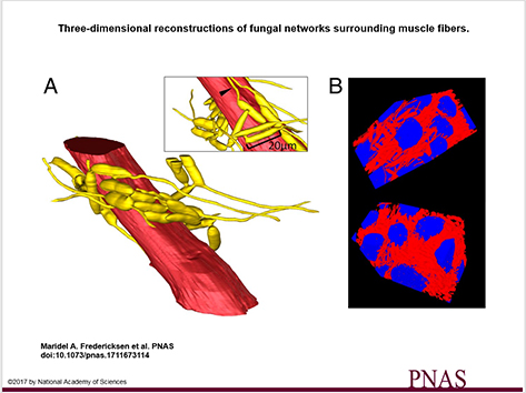
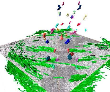
3D visualization and deep-learning reveal complex parasite networks in behaviorally manipulated ants
Microbial parasites may behave collectively to manipulate their host’s behavior. We examine adaptations of a microbial parasite in its natural environment: the body of its coevolved and manipulated host.
Electron microscopy and 3D reconstructions of host and parasite tissues reveal that this fungus invades muscle fibers throughout the ant’s body but leaves the brain intact, and that the fungal cells connect to form extensive networks.
Read more
Maridel A. Fredericksena, Yizhe Zhangb, Missy L. Hazenc, Raquel G. Loretoa,d, Colleen A. Mangoldd,e, Danny Z. Chenb, and David P. Hughes, Department of Entomology, Pennsylvania State University
The importance of context in regulation of gene expression is now an accepted principle; yet the mechanism by which the microenvironment communicates with the nucleus and chromatin in healthy tissues is poorly understood. A functional role for nuclear and cytoskeletal architecture is suggested by the phenotypic differences observed between epithelial and mesenchymal cells…
Read more
Danielle M. Jorgens, Jamie L. Inman, Michal Wojcik, Claire Robertson, Hildur Palsdottir, Wen-Ting Tsai, Haina Huang, Alexandre Bruni-Cardoso, Claudia S. López, Mina J. Bissell, Ke Xu, Manfred Auer
Determining the bacterial cell biology of planctomycetes
Bacteria of the phylum Planctomycetes have been previously reported to possess several features that are typical of eukaryotes, such as cytosolic compartmentalization and endocytosis-like macromolecule uptake. However, recent evidence points towards a Gram-negative cell plan for Planctomycetes, although in-depth experimental analysis has been hampered by insufficient genetic tools…
Read more
Christian Boedeker, Margarete Schüler, Greta Reintjes, Olga Jeske, Muriel C. F. van Teeseling et al.
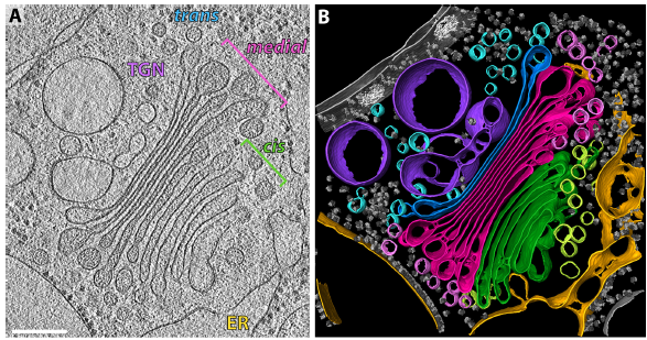
The structure of the COPI coat determined within the cell
COPI-coated vesicles mediate trafficking within the Golgi apparatus and from the Golgi to the endoplasmic reticulum. Here, we applied cryo-focused ion beam milling, cryo-electron tomography and subtomogram averaging to determine the native structure of the COPI coat within vitrified Chlamydomonas reinhardtii cells. The native algal structure resembles the in vitro mammalian structure, but additionally reveals cargo bound beneath beta’–COP. We find that all coat components disassemble... Read more
Yury S Bykov, Miroslava Schaffer, Svetlana O Dodonova, Sahradha Albert, Jurgen M Plitzko, Wolfgang Baumeister, Benjamin D Engel, John AG Briggs
During the acts of biting and chewing, the muscles of the jaw (consisting of the masseter, temporalis, and medial pterygoid in the elevator group, and lateral pterygoid as the main depressor) generate forces that dictate jaw kinematics . The movement of jaws hinges about the temporomandibular joint and are brought together by the muscles attached to respective bones through bone-tendon interfaces known as entheses . Thus, chewing forces affect aspects of craniofacial structure as well as bo... Read more
Kyle H.-Y. Chan, fourth-year undergraduate student in Molecular and Cell Biology, and Public Health at UC Berkeley, FeiFei Yang, Ph.D., postdoctoral scholar in the Laboratory of Multiscale Biomechanics and Biomineralization, School of Dentistry, UCSF, and Sunita P. Ho, Ph.D., Division of Biomaterials and Bioengineering, Department of Preventive and Restorative Dental Sciences, School of Dentistry, University of California San Francisco
3D Reconstruction of Lipid Droplets in the Seed of Brassica napus
Rapeseed is one of the most important and widely cultured oilseed crops for food and nonfood purposes worldwide. Neutral lipids are stored in lipid droplets (LDs) as fuel for germination and subsequent seedling growth. Most of the LD detection in seeds was still in 2D levels, and some of the details might have been lost in previous studies. In the present work, the configuration of LDs in seeds was obtained by confocal imaging combined with 3D reconstruction technology in Brassica napus<... Read more
Yongtai Yin, Liangxing Guo, Kang Chen, Zhenyi Guo, Hongbo Chao, Baoshan Wang, and Maoteng Licorresponding author
Root-knot nematodes induce galls that contain giant-feeding cells harboring multiple enlarged nuclei within the roots of host plants. It is recognized that the cell cycle plays an essential role in the set-up of a peculiar nuclear organization that seemingly steers nematode feeding site induction and development. Functional studies of a large set of cell cycle genes in transgenic lines of the model host Arabidopsis thaliana have contributed to better understand the role of the cell cycle comp... Read more
Antonino de Souza Junior José Dijair, Pierre Olivier, Coelho Roberta R., Grossi-de-Sa Maria F., Engler Gilbert, de Almeida Engler Janice / Institut National de la Recherche Agronomique, Université Côte d’Azur, Centre National de la Recherche Scientifique, Institut Sophia Agrobiotech, Sophia-Antipolis, France